Female reproductive anatomy and physiology
In brief
The female reproductive system includes internal organs in the pelvis (uterus, fallopian tubes, ovaries and vagina) and external organs, together called the vulva. It is supported by the pelvic floor muscles and controlled by hormones produced by the brain and the ovaries. According to FIGO, a normal cycle lasts 24 to 38 days, with periods of no more than 8 days; periods outside these limits warrant medical advice.
Where are the organs of the female reproductive system?
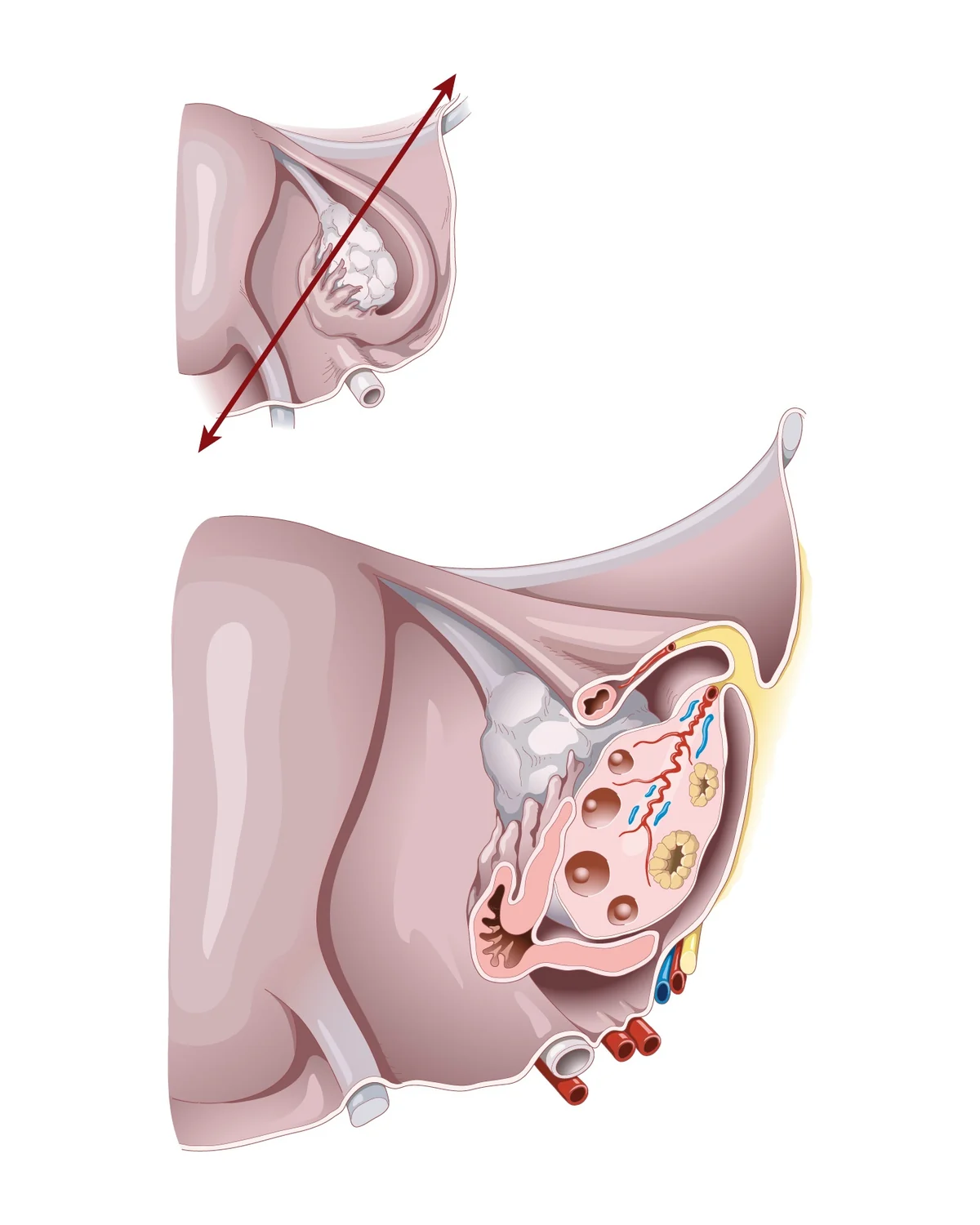
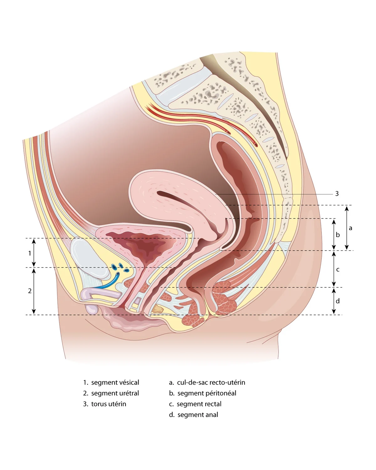
The internal reproductive organs lie in the pelvis, between the bladder in front and the rectum behind. The uterus is usually tilted forwards (anteverted) and rests on the bladder. The peritoneum, the membrane lining the abdominal cavity, covers these organs and forms two pouches: the vesico-uterine pouch in front and the recto-uterine pouch (pouch of Douglas) behind.
This posterior pouch is the lowest point of the abdominal cavity. Fluid or blood collects there, and it is a common site of endometriosis.
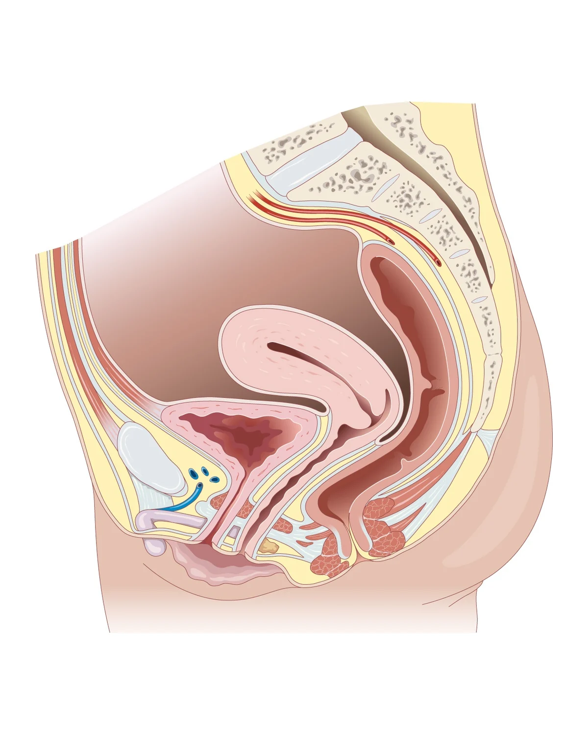
The uterus, fallopian tubes and ovaries
The uterus
The uterus is a hollow, pear-shaped muscle about 7 to 8 cm long outside pregnancy. It has a body, where a pregnancy develops, and a cervix, which opens into the vagina and is examined during a smear test. Its wall has three layers: the endometrium, the lining renewed each cycle; the myometrium, the thick muscle where fibroids arise; and the serosa, which covers it on the outside.
The fallopian tubes
Each tube is about 10 to 12 cm long. It runs from the corner of the uterus and ends near the ovary in a fringed opening that picks up the egg at ovulation. Fertilisation usually takes place in the widest part of the tube, the ampulla.
The ovaries
The two ovaries, about 3 to 4 cm each, produce eggs and female hormones. Each lies in a hollow of the pelvic wall, the ovarian fossa, close to the iliac vessels and the ureter: this is why surgery for an ovarian cyst or endometriosis requires great care.
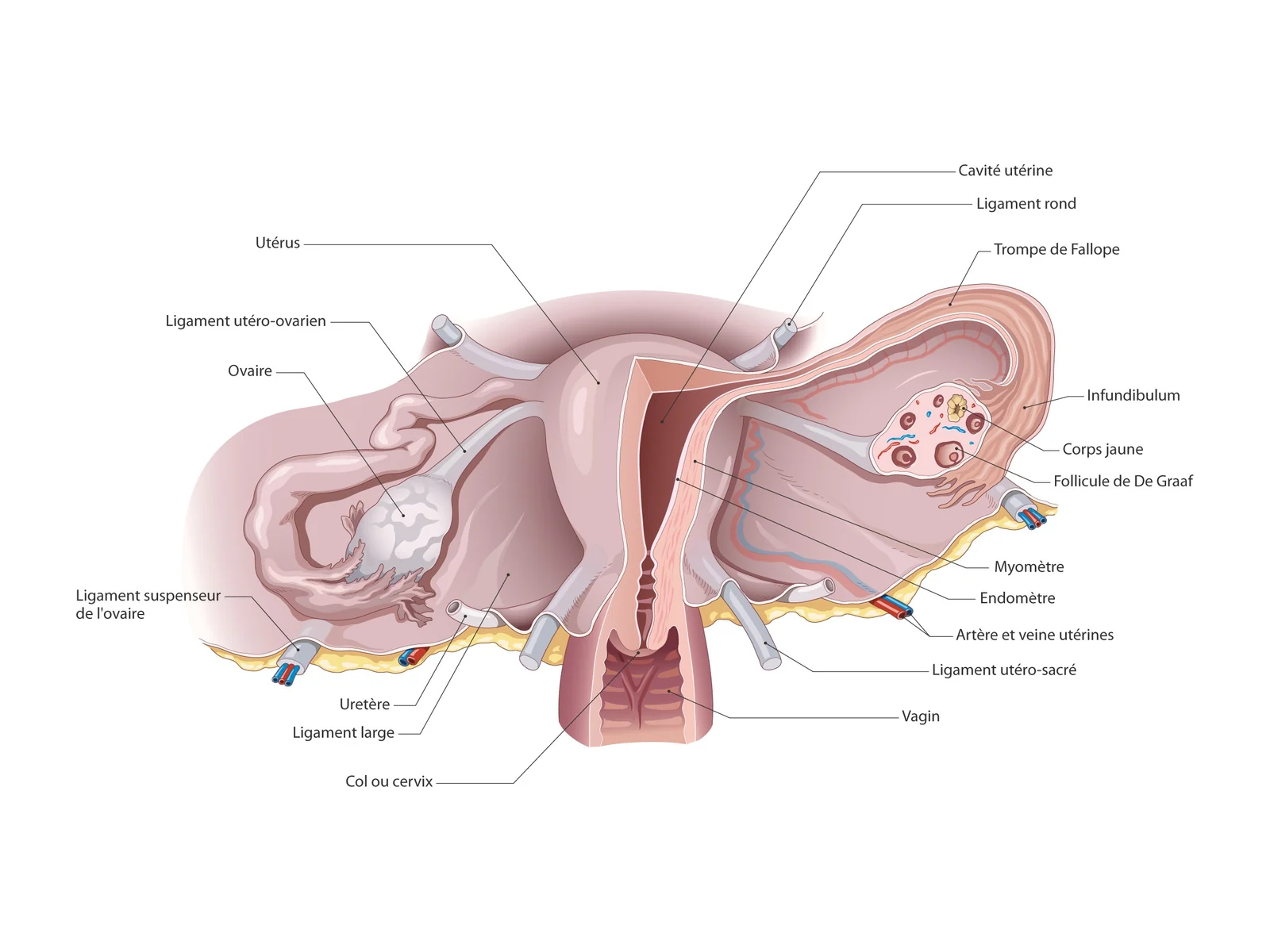
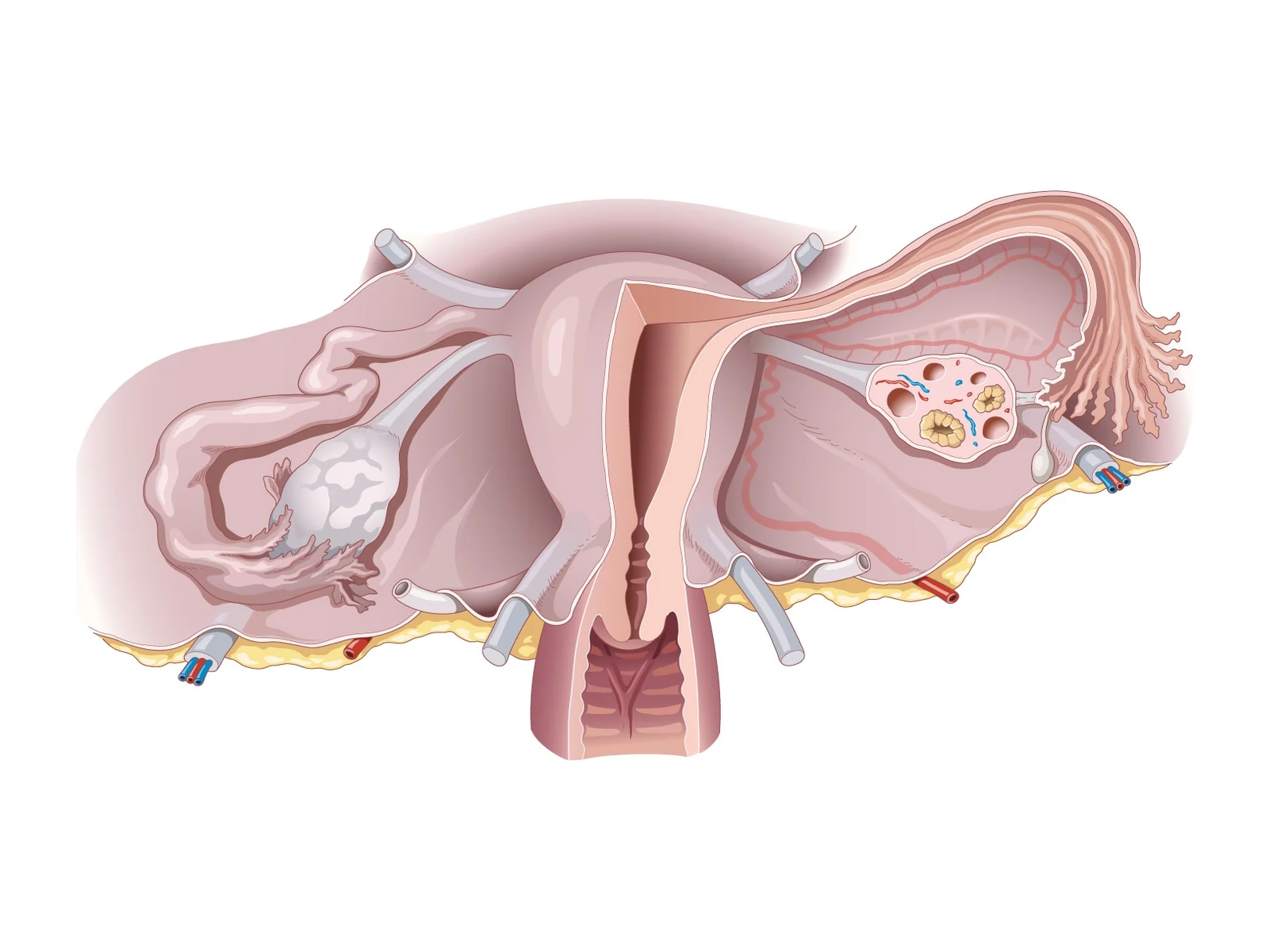
The ligaments
These organs are held by ligaments: the broad ligament, a fold of peritoneum containing the blood vessels; the round ligaments, in front; the uterosacral ligaments, behind, which anchor the cervix to the sacrum; and the suspensory ligament of the ovary, which carries the ovarian vessels. The uterine artery crosses the ureter close to the cervix, a landmark the surgeon identifies during a hysterectomy.
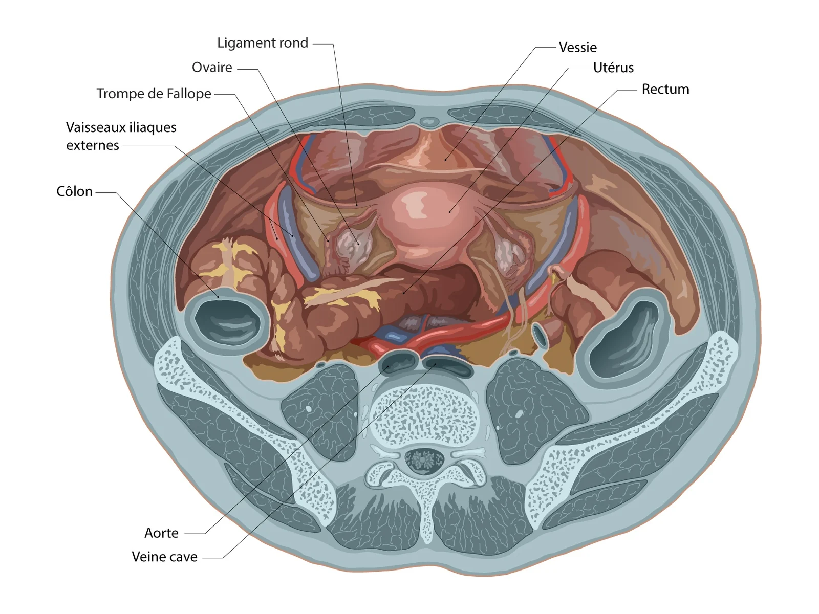
Neighbouring organs
Seen from above, the uterus and adnexa sit in the centre of the pelvis, surrounded by the bladder, rectum, sigmoid colon and the large iliac vessels. These close relations explain why some gynaecological conditions cause urinary or bowel symptoms, and why imaging (pelvic ultrasound, MRI) often comes before surgery.
The vagina
The vagina is a muscular, elastic canal about 7 to 10 cm long that connects the cervix to the vulva. Its upper part surrounds the cervix and forms the vaginal fornices. In front it lies against the bladder and urethra; behind, against the pouch of Douglas, then the rectum and anal canal. These relations matter for understanding pain during sex or prolapse.
The vulva, clitoris and hymen
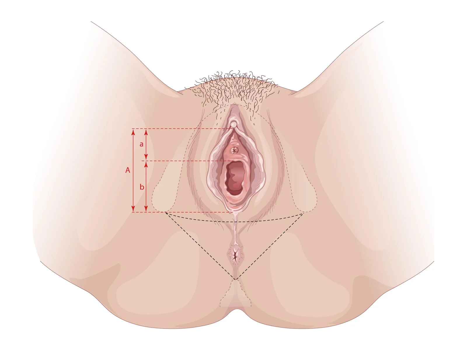
The vulva is the set of external genital organs: the mons pubis, the labia majora and minora, the clitoris, the vestibule where the urethra and vagina open, and the Bartholin's glands. The size and shape of the labia minora vary widely from one woman to another, and this is normal.
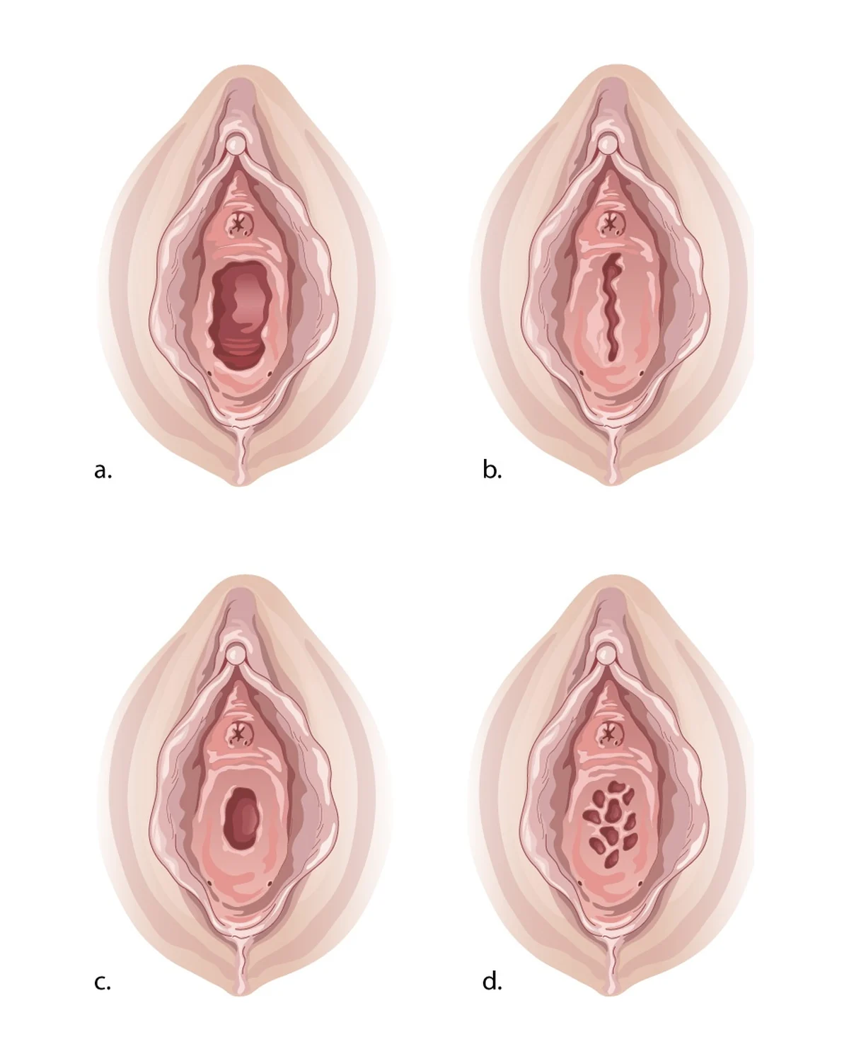
The clitoris is much larger than its visible part, the glans: it has a body, two crura attached to the pelvic bones and two bulbs on either side of the vestibule. The hymen is a thin membrane at the vaginal opening, whose appearance varies between women. A rare imperforate hymen blocks menstrual flow at puberty.
The pelvic floor: a supporting hammock
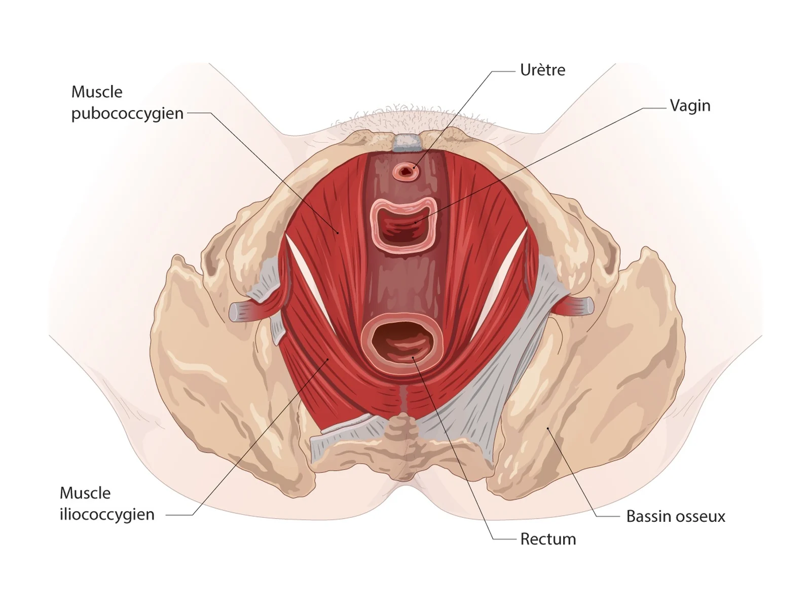
The pelvic floor is a group of muscles, mainly the levator ani, stretched like a hammock beneath the pelvic organs. The urethra, vagina and rectum pass through it. It supports the bladder, uterus and rectum, contributes to continence and contracts during effort.
The vagina is supported at three levels: at the top, by attachments to the cervix and ligaments; in the middle, by lateral attachments to the pelvic wall; at the bottom, by fusion with the centre of the perineum. Vaginal birth can stretch these structures, which partly explains urinary leakage after childbirth and prolapse.
How does the cycle work?
The reproductive system is controlled by a hormonal axis: the hypothalamus releases GnRH, which drives the pituitary gland to produce FSH and LH; these act on the ovaries, which produce oestrogen and progesterone. Each month these hormones prepare the endometrium for a possible pregnancy.
| Phase | What happens |
|---|---|
| Follicular phase | Under FSH, a follicle matures and produces oestrogen; the endometrium thickens. |
| Ovulation | The LH surge triggers release of the egg, which is picked up by the tube. |
| Luteal phase | The corpus luteum produces progesterone, which makes the endometrium ready to receive an embryo. |
| Period | Without pregnancy, the hormonal drop causes the endometrium to shed. |
According to FIGO, a normal cycle lasts 24 to 38 days and a period no more than 8 days. The phases and hormones are explained in detail in our article on the menstrual cycle.
How does the reproductive system change through life?
- Puberty: breast and hair development, then the first period; cycles are often irregular in the first years (see puberty in girls).
- Pregnancy: the uterus grows considerably and rises into the abdomen; it returns to its size within a few weeks after birth.
- Menopause: the ovaries stop working, confirmed after 12 months without a period; falling oestrogen can cause vaginal dryness.
Some women have a variation in the shape of the uterus present from birth (uterine anomaly), classified using the ESHRE/ESGE consensus; it is usually found during fertility tests or an ultrasound (see septate uterus).
Frequently asked questions
What is the normal size of the uterus?
Outside pregnancy, the uterus is about 7 to 8 cm long. Its size varies with age, number of pregnancies and menopause; a pelvic ultrasound can measure it.
Is a retroverted uterus abnormal?
No. The uterus usually tilts forwards, but a backward tilt is a common variant that does not prevent pregnancy or a normal birth.
Where does fertilisation take place?
Usually in the fallopian tube, in its wider part called the ampulla. The embryo then travels to the uterine cavity, where it implants in the endometrium.
What does the pelvic floor do?
It supports the bladder, uterus and rectum and contributes to urinary and anal continence and to sexual function. Pelvic floor rehabilitation is offered after childbirth when needed.
What is the difference between the vulva and the vagina?
The vulva is the external, visible genitals: the outer and inner lips (labia), the clitoris and the vestibule, where the urethra and vagina open. The vagina is an internal passage of about 7 to 10 cm that connects the vulva to the cervix.
Where are the ovaries?
On each side of the uterus, against the wall of the pelvis, in a hollow called the ovarian fossa. Each ovary is about 3 to 4 cm long and lies close to the iliac blood vessels and the ureter.
References
- Munro MG, et al. — The two FIGO systems for normal and abnormal uterine bleeding symptoms and classification of causes of abnormal uterine bleeding in the reproductive years: 2018 revisions. Int J Gynaecol Obstet, 2018 — doi:10.1002/ijgo.12666 — PMID 30198563
- Ashton-Miller JA, DeLancey JO — Functional anatomy of the female pelvic floor. Ann N Y Acad Sci, 2007 — doi:10.1196/annals.1389.034 — PMID 17416924
- Grimbizis GF, et al. — The ESHRE/ESGE consensus on the classification of female genital tract congenital anomalies. Hum Reprod, 2013 — doi:10.1093/humrep/det098 — PMID 23771171

Dr Olivier KadochObstetrician-gynaecologist and gynaecological surgeon in Paris (16th and 5th arrondissements). Former chef de clinique-assistant (senior registrar) at the Paris public hospitals (AP-HP); individually accredited by the French National Authority for Health (HAS). RPPS 10003957809. Background and practice locations
A question about your situation?Appointments at the practice or by video consultation, usually within one week to 10 days.
Book an appointmentGeneral information article: it does not replace a consultation. In an emergency, call 15 or 112 in France, or your local emergency number.