Endometrial (womb) cancer: symptoms, diagnosis and treatment
In brief
In more than 9 out of 10 cases, endometrial cancer (cancer of the body of the womb) first shows itself through abnormal vaginal bleeding, most often after the menopause. Any bleeding after the menopause, however slight, needs a prompt appointment for an ultrasound and, if needed, an endometrial biopsy. Found early, it is often cured by surgery to remove the uterus, tubes and ovaries, sometimes followed by radiotherapy or other treatments decided at a multidisciplinary team meeting.
What is endometrial cancer?
Cancer of the body of the uterus (womb) starts in the vast majority of cases in the endometrium, the lining of the inside of the uterus that renews itself with each cycle. This is called endometrial carcinoma. It is different from cervical cancer, which has other causes (human papillomavirus) and its own screening. Uterine sarcomas, which arise from the muscle, are much rarer.
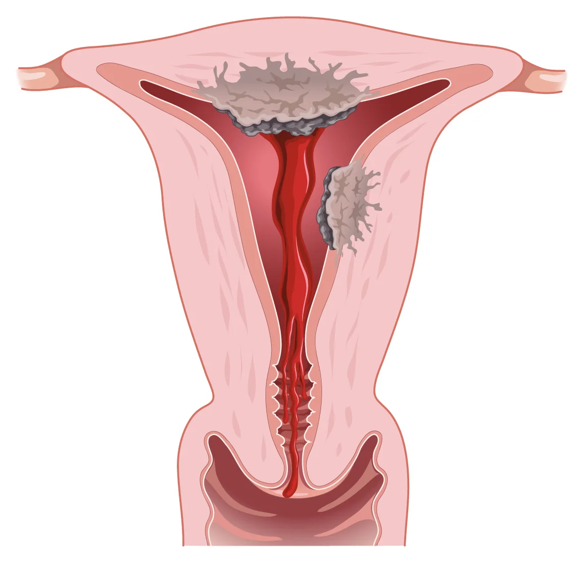
It is the most common pelvic gynaecological cancer in Western countries. It mainly affects postmenopausal women, but it can occur earlier, particularly when there is a genetic predisposition. For an overview of gynaecological cancers, see our Gynaecological cancer care page.
What are the warning signs of endometrial cancer?
The main sign is abnormal vaginal bleeding. A large meta-analysis shows that 91% of women with endometrial cancer had bleeding after the menopause. Conversely, about 9% of women who bleed after the menopause have cancer: the cause is therefore usually benign, but it must always be checked.
- After the menopause: any bleeding, even a single trace of pink or brownish spotting, needs a prompt appointment.
- Before or around the menopause: periods that have become very heavy or prolonged, bleeding between periods, especially if there are risk factors.
- More rarely: watery or pink discharge, pelvic pain.
Other causes of bleeding after the menopause (thinning of the lining, polyp, hormone treatment) are covered in our article on bleeding after the menopause.
Who is most at risk of endometrial cancer?
Most endometrial cancers are driven by long-term exposure to oestrogen that is not balanced by progesterone:
- being overweight or obese (fatty tissue produces oestrogen);
- never having been pregnant, early puberty, late menopause;
- polycystic ovary syndrome with very infrequent cycles;
- oestrogen-only treatment in a woman who still has her uterus, without an added progestogen;
- long-term tamoxifen treatment after breast cancer;
- diabetes and high blood pressure.
Lynch syndrome, an inherited predisposition, clearly increases the risk of endometrial and bowel cancer. It is suspected when there is a family history of these cancers or of cancer at a young age.
There is no screening programme for endometrial cancer: neither the smear test nor routine ultrasound is recommended for women without symptoms.
Which tests are used to diagnose endometrial cancer?
| Step | Test | Purpose |
|---|---|---|
| 1 | Gynaecological examination | Check that the bleeding comes from the uterus and not from the cervix or vagina. |
| 2 | Transvaginal pelvic ultrasound | Measure the thickness of the endometrium and look for any abnormality. A thin endometrium makes cancer unlikely. |
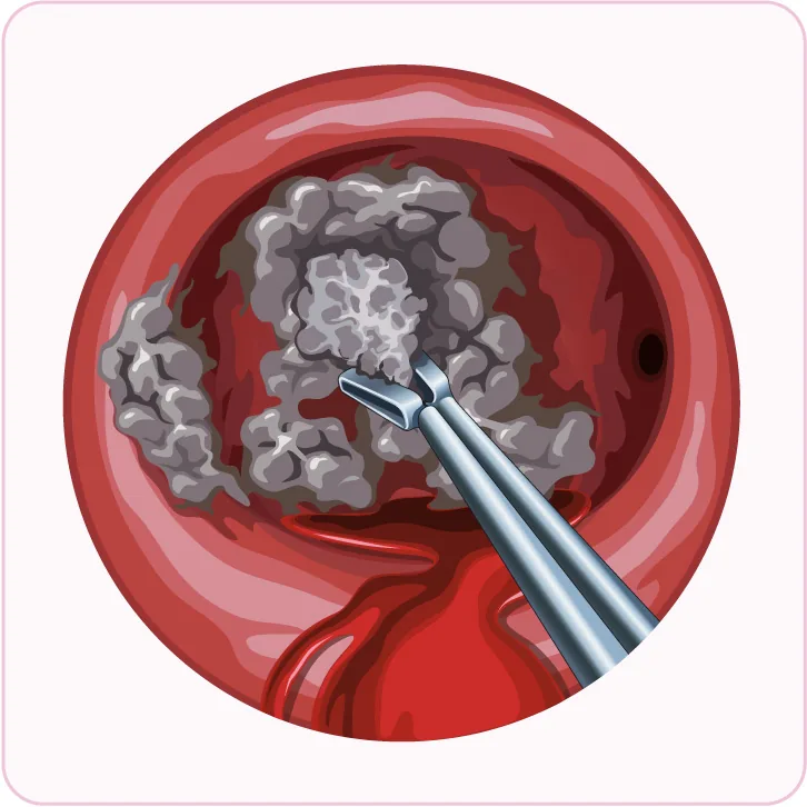
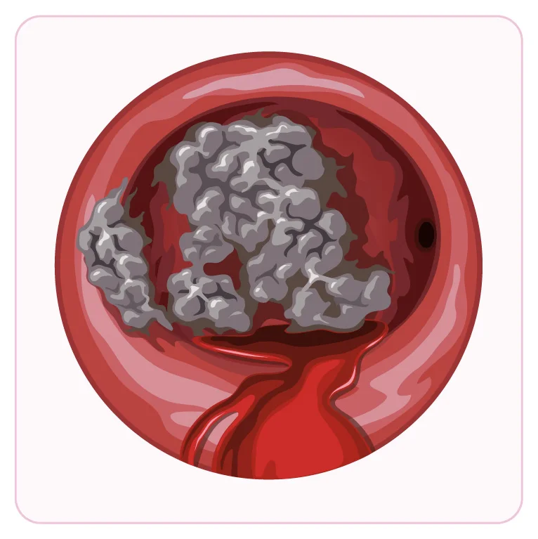
| 3 | Endometrial biopsy (pipelle sampling in clinic) or hysteroscopy with targeted biopsies | Obtain a definite diagnosis under the microscope, with the type and grade of the tumour. |
| 4 | Molecular analysis of the tumour | Classify the tumour into 4 groups (POLE, MMR deficiency, abnormal p53, no specific molecular profile): this refines the outlook and treatment. |
| 5 | Pelvic MRI, plus CT scan of the chest, abdomen and pelvis depending on the type | Assess invasion of the uterine muscle, involvement of the cervix and lymph nodes, and distant spread. |
Testing the tumour for a deficiency in DNA mismatch repair (MMR) proteins is recommended for all patients. It guides treatment and is used to screen for Lynch syndrome.
How is endometrial cancer treated?
The strategy is decided at a multidisciplinary team meeting (MDT), based on the FIGO stage, the tumour type, the molecular profile, your general health and your wishes.
Surgery
This is the main treatment for localised disease: total hysterectomy with removal of both fallopian tubes and both ovaries. A minimally invasive approach (laparoscopy) is preferred when the disease is confined to the uterus, because it reduces complications and hospital stay. Lymph nodes are now usually assessed using the sentinel lymph node technique, which identifies and removes only the first draining nodes. In most cases this replaces full lymph node dissection and reduces the risk of leg lymphoedema. To learn more about the operation, see our article on hysterectomy.
Additional treatments
- No further treatment for low-risk disease.
- Vaginal vault brachytherapy: internal radiotherapy to lower the risk of local recurrence in intermediate-risk disease.
- External pelvic radiotherapy, sometimes combined with chemotherapy, for high-risk disease or when lymph nodes are involved.
- Chemotherapy and, for advanced or recurrent disease, immunotherapy, particularly when the tumour is MMR-deficient.
Special situations
In a young woman who wants to become pregnant and whose tumour is low grade with no muscle invasion, fertility-sparing treatment with high-dose progestogens and regular hysteroscopic checks may be considered in a specialist centre. For a patient who cannot have surgery, radiotherapy or hormone therapy are alternatives.
What is the outlook for endometrial cancer?
Endometrial cancer is often found at an early stage, because bleeding appears early. In that case the outlook is generally good. It depends mainly on the stage, the tumour type, whether tumour cells are found in the blood or lymph vessels, and the molecular group.
After treatment, regular follow-up is arranged: see our article on follow-up after uterine cancer.
What about life after treatment?
Most women return to normal life a few weeks after minimally invasive surgery. Removing the ovaries brings on the menopause if it had not yet occurred; its symptoms can be treated. Regular physical activity and keeping weight, blood pressure and diabetes under control are good for overall health. If Lynch syndrome is confirmed, a cancer genetics consultation is offered, as your relatives can benefit from appropriate screening.
When should you see a doctor urgently?
- Any bleeding after the menopause: book an appointment within a few days, without waiting for your annual check-up.
- Heavy bleeding (soaking more than one pad an hour for several hours), feeling faint, pallor, breathlessness: go to A&E or, in France, call 15 (or 112), or your local emergency number.
- After surgery: fever, significant calf pain, sudden breathlessness, worsening abdominal pain: contact the surgical team or, in France, call 15.
Would you like a medical opinion?Appointments in Paris 16th or 5th, or by video consultation.
Book an appointmentFrequently asked questions
Does a smear test detect endometrial cancer?
No. The smear test and the HPV test are used to screen for cervical cancer. They may sometimes show abnormal cells coming from the endometrium, but a normal smear does not rule out endometrial cancer.
Does a thick endometrium on ultrasound mean cancer?
Not necessarily. A thick endometrium may be due to a polyp, benign hyperplasia or a hormonal effect. In a postmenopausal woman who is bleeding, it usually leads to a biopsy to reach a diagnosis.
Does menopausal hormone therapy increase the risk?
Oestrogen taken alone increases the risk in a woman who still has her uterus. That is why a progestogen is always added in this case, to protect the endometrium.
Do the ovaries need to be removed after the menopause?
In endometrial cancer, removing the ovaries and fallopian tubes is part of standard treatment. In a young woman with early, low-grade disease, keeping the ovaries may be considered.
How long is the hospital stay after the operation?
As a guide, after a laparoscopic (keyhole) hysterectomy the stay is generally 2 to 4 days and time off work about a month, with no heavy lifting for 6 weeks. These are adjusted to each patient and to any further treatment. At the practice, surgery for gynaecological cancers is carried out at the Clinique Ambroise Paré (Neuilly-sur-Seine); a written estimate of fees is given before the operation.
References
- Concin N, et al. ESGO-ESTRO-ESP guidelines for the management of patients with endometrial carcinoma: update 2025. The Lancet Oncology, 2025. doi:10.1016/S1470-2045(25)00167-6 — PMID 40744042
- Clarke MA, et al. Association of Endometrial Cancer Risk With Postmenopausal Bleeding in Women: A Systematic Review and Meta-analysis. JAMA Internal Medicine, 2018. doi:10.1001/jamainternmed.2018.2820 — PMID 30083701
- Poudou C, et al. Balance bénéfice-risque du traitement hormonal de la ménopause : cancers et mortalité. Recommandations pour la pratique clinique du CNGOF et du GEMVi. [Benefit-risk balance of hormone replacement therapy: cancers and mortality. CNGOF and GEMVi guidelines] Gynécologie Obstétrique Fertilité & Sénologie, 2021. doi:10.1016/j.gofs.2021.03.031 — PMID 33771739

Dr Olivier KadochObstetrician-gynaecologist and gynaecological surgeon in Paris, seeing patients in Paris 16th and at the Centre de la Femme Paris 5. Former chef de clinique-assistant (senior registrar) at the Paris public hospitals (AP-HP); individually accredited by the French National Authority for Health (HAS). RPPS 10003957809. Background and practice locations
A question about your situation?Appointments at the practice or by video consultation, usually within one week to 10 days.
Book an appointmentGeneral information article: it does not replace a medical consultation. In an emergency, call 15 or 112 in France, or your local emergency number.