Diagnostic hysteroscopy: how it works, when it is used, what happens
In brief
Diagnostic hysteroscopy means looking directly inside the uterus with a small camera passed through the vagina and cervix, without any incision. Usually performed in the consultation room without anaesthetic, it takes a few minutes and is used to investigate abnormal bleeding, infertility or recurrent miscarriage. It also allows targeted biopsies.
Dedicated website. Everything about diagnostic and operative hysteroscopy: hysteroscopie-paris.fr.
What is a diagnostic hysteroscopy?
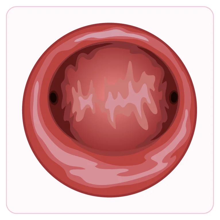
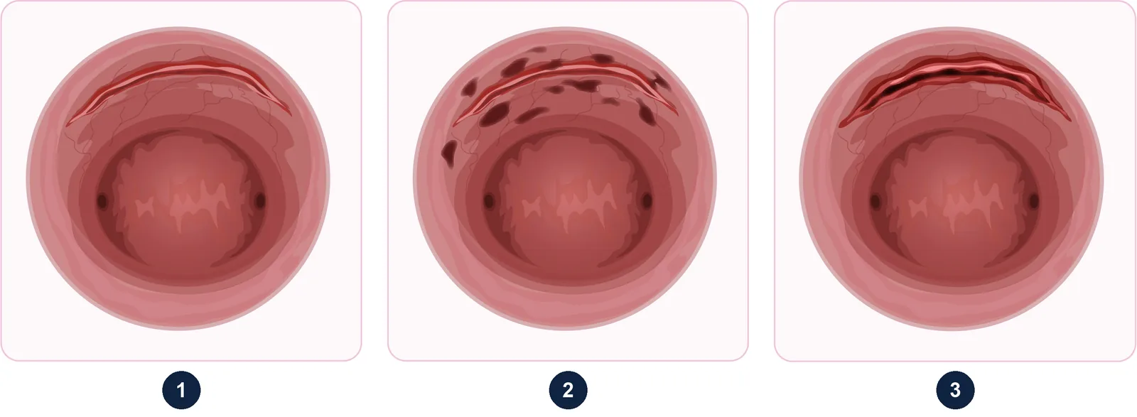
A hysteroscope is a very thin optical tube connected to a light source and a camera. It is passed through the vagina and then the cervix into the cavity of the uterus, which is gently opened up with saline (salt water). The image appears on a screen: the doctor examines the endometrium (the lining of the uterus), the shape of the cavity and the openings of the fallopian tubes.
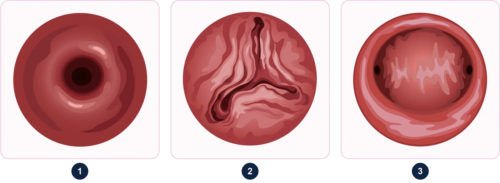
There are two types of hysteroscopy:
- diagnostic hysteroscopy, the subject of this article: a visual examination, sometimes with a biopsy;
- operative hysteroscopy, which treats lesions (polyps, fibroids, septa, adhesions), usually in the operating theatre. It is described on the Operative hysteroscopy page.
For outpatient (office) hysteroscopy, French guidelines from the French College of Gynaecologists and Obstetricians (CNGOF) recommend the vaginoscopic technique (no speculum and no instrument holding the cervix), with a small-diameter hysteroscope (sheath of 3.5 mm or less) and saline, and without routine anaesthetic, cervical preparation, antibiotics or vaginal disinfection.
Why would I need a hysteroscopy?
The main indications are:
- Abnormal bleeding: heavy or prolonged periods, bleeding between periods and above all bleeding after the menopause. Hysteroscopy can show a polyp, a submucosal fibroid or hyperplasia, and allows suspicious areas to be biopsied.
- Fertility investigations: looking for abnormalities that could interfere with embryo implantation (polyp, fibroid, adhesions, septum), particularly before in vitro fertilisation.
- Recurrent miscarriage: checking that the cavity is normal.
- An abnormal finding on ultrasound or saline infusion sonography that needs confirming.
- A check-up after surgery on the cavity or a D&C (curettage), to look for adhesions.
- More rarely, locating a coil (IUD) or a fragment of tissue left in the uterus.
According to a European evidence-based guide, office hysteroscopy is the most accurate test for diagnosing precancerous and cancerous lesions of the uterine cavity; any lesion removed must be examined under the microscope.
Who should not have a hysteroscopy?
- Current or possible pregnancy: the procedure could end it. If in doubt, a pregnancy test is done first.
- An untreated genital infection (endometritis, salpingitis, pelvic infection), which the procedure could worsen or spread.
- Heavy bleeding at the time, which obscures the view; the procedure is then postponed.
Suspected endometrial cancer is not a contraindication: according to the CNGOF, hysteroscopy can still be performed in this case.
How do I prepare for a diagnostic hysteroscopy?
- Timing in your cycle: if you still have periods, the procedure is ideally scheduled after your period and before ovulation, when the lining is thin and pregnancy is unlikely. After the menopause, it can be done at any time.
- Tell the doctor about a possible pregnancy, unusual discharge, fever or recent pelvic pain.
- Pain: a 2020 meta-analysis showed that an anti-inflammatory taken by mouth beforehand reduces pain during and after the procedure. Your doctor will tell you whether you can take one, depending on your medical history.
- No need to fast for an outpatient procedure without anaesthetic.
- Your medicines: mention any anticoagulants or antiplatelet drugs.
- Practical arrangements: the procedure is performed at the practice; appointments can be booked via Doctolib or by calling 01 43 79 59 79 (the average waiting time for an appointment is one week to 10 days, except during holiday periods); fees and reimbursement are explained to you at the consultation.
What happens during a diagnostic hysteroscopy?
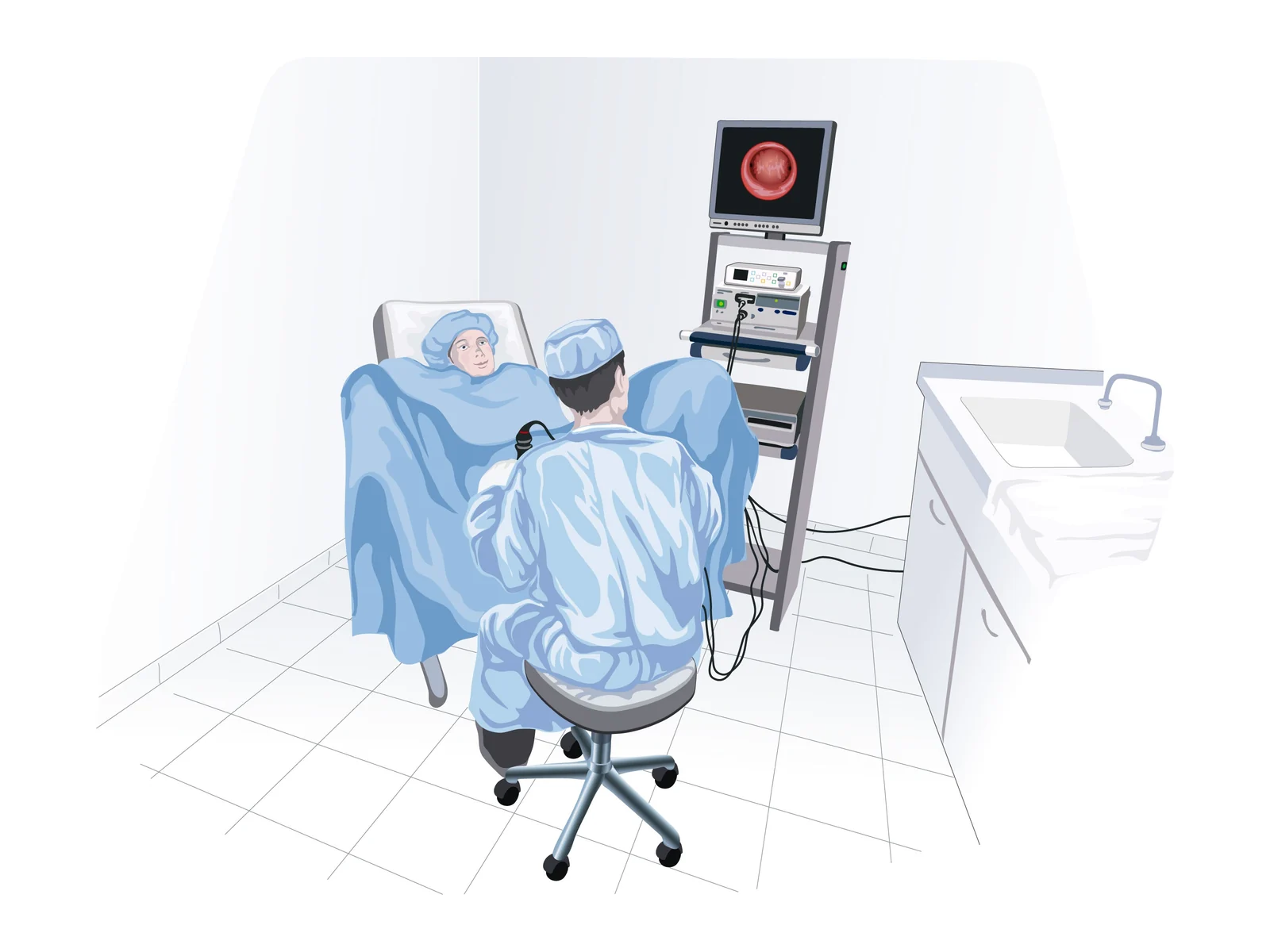
- You lie in the gynaecological examination position.
- The hysteroscope is gently passed into the vagina and through the cervix, usually without a speculum.
- Saline opens up the cavity; the doctor examines it systematically and you can follow the images on the screen.
- If needed, a biopsy of the lining is taken.
- The hysteroscope is removed; the fluid drains out through the vagina.
The procedure takes a few minutes. The most common sensation is a cramp similar to period pain, when the scope passes through the cervix and the cavity is distended. Report any severe pain: the doctor can slow down or stop. Feeling briefly faint is possible; you will be given a few minutes to rest before leaving.
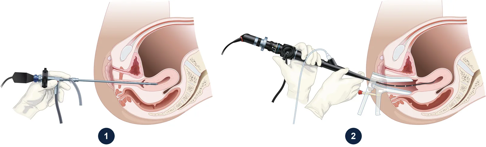
If the procedure cannot be done in the consultation room (very tight cervix, too much pain, anxiety), it can be performed under anaesthetic as a day case, often combined with a surgical procedure.
What can a hysteroscopy find?
| Finding | What it means | Possible next step |
|---|---|---|
| Normal cavity | No cause within the uterus | Look for other causes depending on the situation |
| Polyp | A growth of the lining, usually benign | Removal by operative hysteroscopy and analysis |
| Submucosal fibroid | A benign muscle tumour bulging into the cavity | Hysteroscopic resection depending on size and symptoms |
| Adhesions (synechiae) | Scar tissue joining the walls | Hysteroscopic division |
| Uterine septum | A malformation | Hysteroscopic division in some situations |
| Thickened or irregular lining | Hyperplasia or, more rarely, cancer | Biopsy, pathology result |
What the doctor has seen is explained to you at the end of the procedure. The result of a biopsy, examined under the microscope, comes later and is sent to you via Doctolib or by post (by phone if it is urgent).
Why hysteroscopy rather than another test?
Investigation of the uterine cavity generally follows a step-by-step approach, from the simplest to the most complete:
- the pelvic ultrasound, the first-line test;
- saline infusion sonography, which outlines the cavity more clearly when the ultrasound is uncertain;
- diagnostic hysteroscopy, which allows direct viewing and biopsy exactly where needed.
Hysteroscopy has replaced "blind" curettage (D&C) for diagnosis. A European evidence-based guide on endometrial polyps points out that curettage without visual control should be avoided, as it misses localised lesions. The same guide recommends, for a postmenopausal woman with bleeding and a suspected polyp, hysteroscopy with removal of the polyp and microscopic analysis.
Hysteroscopy gives no information about the ovaries or the thickness of the uterine muscle: it is therefore often combined with an ultrasound, or even an MRI for large fibroids or adenomyosis. Nor does it replace cervical screening (smear tests).
What is recovery like and when should I seek help?
You can return to your usual activities the same day. Watery discharge and light bleeding for one to two days, along with mild cramps, are normal.
Complications of diagnostic hysteroscopy are rare: fainting (vasovagal reaction), infection, and very rarely perforation of the uterus. See a doctor promptly if you have:
- fever or chills;
- lower abdominal pain that is getting worse;
- unpleasant-smelling discharge;
- heavy bleeding.
If you feel very faint or have severe pain, call 15 or 112 in France, or your local emergency number.
Would you like a medical opinion?Appointments in Paris 16th or 5th, or by video consultation.
Book an appointmentFrequently asked questions
Is a diagnostic hysteroscopy painful?
Most women describe a brief cramp similar to period pain. Small-diameter scopes and the speculum-free technique limit discomfort; an anti-inflammatory taken beforehand can reduce pain if your doctor agrees.
Do I need a general anaesthetic for a hysteroscopy?
No, as a general rule. Diagnostic hysteroscopy is done in the consultation room without anaesthetic. Anaesthesia is only considered if the procedure is not possible this way or is combined with surgery.
Can I have a hysteroscopy with a coil (IUD) in place?
Yes, it is possible, and it is even a way to locate a coil that is difficult to remove. Mention it before the procedure.
When can I exercise, have sex and take baths again after a hysteroscopy?
As a guide, exercise can generally be resumed straight away, except swimming. It is generally advised to wait about 15 days before having sex, taking baths, swimming or using tampons. This guidance is tailored to your situation at the consultation, especially if a biopsy was taken.
How much does a diagnostic hysteroscopy cost?
You are told the fee before the procedure. At the practice, students are seen at the national health insurance rate, and patients covered by the Complémentaire santé solidaire (C2S) or AME schemes are accepted with no extra fees.
References
- Deffieux X, et al. Prevention of the complications related to hysteroscopy: guidelines for clinical practice (CNGOF). J Gynecol Obstet Biol Reprod (Paris), 2013. doi:10.1016/j.jgyn.2013.09.008 — PMID 24210234
- Vitale SG, et al. Endometrial polyps. An evidence-based diagnosis and management guide. Eur J Obstet Gynecol Reprod Biol, 2021. doi:10.1016/j.ejogrb.2021.03.017 — PMID 33756339
- De Silva PM, et al. Analgesia for office hysteroscopy: a systematic review and meta-analysis. J Minim Invasive Gynecol, 2020. doi:10.1016/j.jmig.2020.01.008 — PMID 31982584

Dr Olivier KadochObstetrician-gynaecologist and gynaecological surgeon in Paris, seeing patients in Paris 16th and at the Centre de la Femme Paris 5. Former chef de clinique-assistant (senior registrar) at the Paris public hospitals (AP-HP); individually accredited by the French National Authority for Health (HAS). RPPS 10003957809. Background and practice locations
A question about your situation?Appointments at the practice or by video consultation, usually within one week to 10 days.
Book an appointmentGeneral information article: it does not replace a medical consultation. In an emergency, call 15 or 112 in France, or your local emergency number.