Myomectomy: removing a fibroid while preserving the uterus
In brief
Myomectomy is the operation that removes one or more fibroids (myomas) while leaving the uterus in place, which preserves the possibility of a pregnancy. The surgical approach depends mainly on the position of the fibroid according to the FIGO classification: hysteroscopy for fibroids that protrude into the cavity (types 0 to 2), laparoscopy or laparotomy for the others. A Cochrane review of 9 randomised trials (808 women) shows that with laparoscopy, post-operative pain, fever and length of hospital stay are lower than with open surgery, with no difference in the risk of recurrence.
What is a myomectomy, and who is it for?
A fibroid is a benign tumour of the uterine muscle. Myomectomy involves removing it while preserving the rest of the uterus, unlike hysterectomy, which removes the whole uterus.
It is intended for women who wish to keep their uterus, particularly if they are planning a pregnancy, and who have:
- symptoms related to the fibroid (heavy periods, anaemia, pain, pressure on the bladder or rectum);
- or a fibroid distorting the uterine cavity associated with infertility or miscarriages.
A fibroid with no symptoms and no effect on fertility generally does not need surgery. When treatment is needed is explained on the Fibroids page.
What is the FIGO classification for?
The International Federation of Gynecology and Obstetrics (FIGO) has defined a classification of fibroids into 9 types (0 to 8) according to their relationship with the uterine cavity and the uterine wall, revised in 2018. It guides the choice of surgical approach.
| FIGO type | Location | Usual approach |
|---|---|---|
| 0 | Entirely within the cavity (pedunculated) | Hysteroscopy |
| 1 | Within the cavity, less than 50% in the muscle | Hysteroscopy |
| 2 | Within the cavity, 50% or more in the muscle | Hysteroscopy, sometimes in 2 stages |
| 3 | In contact with the endometrium, without protruding into the cavity | Laparoscopy or laparotomy |
| 4 | Entirely within the thickness of the muscle (intramural) | Laparoscopy or laparotomy |
| 5 – 6 | Beneath the serosa, more or less than 50% in the muscle | Laparoscopy or laparotomy |
| 7 | Pedunculated subserosal | Laparoscopy |
| 8 | Other (cervix, broad ligament, etc.) | Case by case |
The pre-operative assessment is based on pelvic ultrasound, often supplemented by saline infusion sonography or diagnostic hysteroscopy for fibroids close to the cavity, and by pelvic MRI in the case of multiple or large fibroids. These tests specify the number, size and FIGO type of each fibroid and the thickness of muscle remaining between the fibroid and the outer surface of the uterus.
How is a hysteroscopic myomectomy performed?
Operative hysteroscopy goes through the natural passages: a thin camera is inserted through the cervix into the uterine cavity, which is distended with fluid. The fibroid is cut into chips (resection) or removed with an intrauterine morcellation system, without any skin incision.
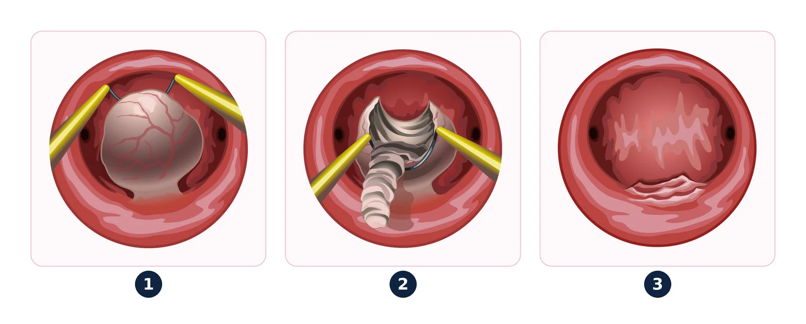
Key points:
- the procedure is generally performed as a day case, under general or regional anaesthesia;
- a large type 2 fibroid may require two operations;
- the main risks are uterine perforation, fluid overload (distension fluid passing into the bloodstream), infection and, later on, adhesions inside the cavity (synechiae).
The 2022 guidelines of the CNGOF (French College of Gynaecologists and Obstetricians) on abnormal uterine bleeding devote a chapter to type 0 to 2 fibroids causing bleeding, for which hysteroscopic resection is the recommended surgical treatment.
Laparoscopy or laparotomy: how is the choice made?
For type 3 to 7 fibroids, the uterus must be incised from the outside. Two approaches are possible.
Laparoscopy (minimally invasive surgery) uses a camera and instruments inserted through 3 or 4 small incisions in the abdomen. The fibroid is enucleated, then the uterus is stitched in several layers.
Laparotomy is a conventional opening of the abdomen, most often horizontal above the pubic bone. It remains useful for very numerous, very large or deeply situated fibroids, for which a strong repair of the uterus is difficult by laparoscopy.
According to the Cochrane review by Bhave Chittawar et al. (2014), laparoscopy, compared with open surgery, is associated with:
- less post-operative pain at 6 and 48 hours;
- a risk of post-operative fever roughly halved (odds ratio 0.44);
- a shorter hospital stay;
- no demonstrated difference in fibroid recurrence.
However, data are lacking to compare rare risks precisely (uterine rupture in a later pregnancy, thrombosis). The choice therefore takes into account the number, size and location of the fibroids, and the expected quality of the uterine repair.
What is morcellation, and is it a problem?
In laparoscopy, a fibroid several centimetres across cannot come out as it is through a 1 cm incision. It has to be broken into fragments: this is morcellation. It can be performed:
- with a power morcellator;
- or in a "contained" way, inside a sealed bag introduced into the abdomen, to prevent fragments from scattering;
- or through a short additional incision (mini-laparotomy) or through the vagina.
The main concern is the rare risk that an apparently benign tumour is in fact a uterine sarcoma (malignant tumour of the muscle). Uncontained morcellation could then spread malignant cells in the abdomen. This is why pre-operative imaging (MRI if in doubt), age, menopause and rapid growth are taken into account before morcellation is proposed, and why in-bag morcellation is increasingly used.
What is the risk of bleeding, and how is it limited?
A fibroid is a tumour with a very rich blood supply: bleeding is the main risk of myomectomy, particularly for large, multiple or intramural fibroids.
Several measures can reduce it:
- correcting anaemia before the operation (iron, sometimes temporary hormone treatment to stop periods);
- local injection of vasoconstrictors into the uterine muscle during the operation;
- tranexamic acid in some cases;
- in selected situations, temporary control of the uterine arteries.
A blood transfusion may be needed, rarely for a single fibroid, more often for multiple fibroids operated on by laparotomy. Exceptionally, uncontrollable haemorrhage may require a hysterectomy during the operation: this risk is part of the information given before surgery.
What is the recovery like?
These figures are a guide only and are tailored to each patient at the consultation:
| Hysteroscopy | Laparoscopy | Laparotomy | |
|---|---|---|---|
| Incision | None | 3 to 4 incisions of 5 to 10 mm | Incision of 8 to 12 cm |
| Hospital stay | Day case in most cases | 2 to 3 days | 3 to 5 days |
| Sick leave | 2 to 4 days | 2 to 4 weeks | 4 to 6 weeks (desk job), 6 to 8 weeks (physical job) |
| Bleeding after the operation | Pinkish discharge for a few days | Moderate | Moderate |
| Exercise | Straight away, except swimming | 1 to 3 weeks | 6 to 12 weeks |
| Sex | About 15 days | 1 to 2 weeks | 4 to 8 weeks |
| Baths | 15 days | 4 weeks | 4 weeks |
| Effect on a future pregnancy | Generally no scar in the muscle | Uterine scar | Uterine scar |
The operation can be performed at the Hôpital privé Geoffroy Saint-Hilaire (Paris 5th), the Clinique de la Muette (Paris 16th) or the Clinique Ambroise Paré (Neuilly-sur-Seine). The surgical fees are set out in a quote given to you beforehand.
After laparoscopy or laparotomy, activities are resumed gradually. Pain is usually controlled with simple painkillers. A follow-up consultation generally takes place 4 to 6 weeks after the operation, with the report of the microscopic analysis of the fibroid.
Can you get pregnant after a myomectomy?
Yes, this is often the aim of the operation.
When can a pregnancy be considered? After a myomectomy that has opened the uterine muscle (types 3 to 7), a healing period is usually advised before conceiving. The length chosen depends on the extent of the scar: the wait before a new pregnancy is discussed at the post-operative consultation. After a simple hysteroscopic resection, this wait is generally short.
Is a caesarean needed? The uterine scar carries a rare risk of uterine rupture during pregnancy or labour. The mode of delivery is discussed case by case on the basis of the operative report: depth of the scar, whether or not the uterine cavity was opened, number of fibroids removed. It is therefore important to keep this report and pass it on to the team that will follow the pregnancy.
Finally, fibroids can recur after a myomectomy: new fibroids may appear in the following years, hence the value of ultrasound monitoring.
When should I seek urgent care?
After a myomectomy, seek medical advice without delay or call 15 (SAMU) or 112 in France, or your local emergency number, in the event of:
- heavy bleeding (more than one pad per hour);
- fever above 38.5 °C;
- abdominal pain that gets worse instead of better;
- feeling faint, breathlessness, pain or swelling in a calf;
- during a later pregnancy: sudden, severe abdominal pain, especially if you have a known uterine scar.
Frequently asked questions
Does a myomectomy stop fibroids from coming back?
No. It removes the fibroids present, but the uterus keeps its ability to form new ones. The risk of recurrence decreases after menopause, when fibroids are no longer stimulated by hormones.
Can several fibroids be removed in a single operation?
Yes, this is common. The number and location of the fibroids then influence the choice of approach and the risk of bleeding.
Is a myomectomy performed under general anaesthesia?
Usually yes for laparoscopy and laparotomy. Hysteroscopy can sometimes be performed under regional anaesthesia.
Is the fibroid analysed after the operation?
Yes, always. Microscopic examination confirms the benign nature of the fibroid in the vast majority of cases.
Is treatment needed before the operation?
Sometimes, to correct anaemia or reduce the size of the fibroid. This decision is individual and discussed at the pre-operative consultation.
How long is the recovery after a myomectomy?
As a guide, time off work is 2 to 4 days after hysteroscopy, 2 to 4 weeks after laparoscopy and 4 to 8 weeks after laparotomy, depending on your job. Activities are resumed gradually; instructions are tailored to each patient at the consultation.
References
- Munro MG, et al. The two FIGO systems for normal and abnormal uterine bleeding symptoms and classification of causes of abnormal uterine bleeding in the reproductive years: 2018 revisions. Int J Gynaecol Obstet, 2018. doi:10.1002/ijgo.12666 — PMID 30198563
- Bhave Chittawar P, et al. Minimally invasive surgical techniques versus open myomectomy for uterine fibroids. Cochrane Database Syst Rev, 2014. doi:10.1002/14651858.CD004638.pub3 — PMID 25331441
- Brun JL, et al. [Management of women with abnormal uterine bleeding: Clinical practice guidelines of the French National College of Gynecologists and Obstetricians (CNGOF)]. Gynecol Obstet Fertil Senol, 2022. doi:10.1016/j.gofs.2022.02.078 — PMID 35248756

Dr Olivier KadochObstetrician-gynaecologist and gynaecological surgeon in Paris (16th and 5th arrondissements). Former chef de clinique-assistant (senior registrar) at the Paris public hospitals (AP-HP); individually accredited by the French National Authority for Health (HAS). RPPS 10003957809. Background and practice locations
A question about your situation?Appointments at the practice or by video consultation, usually within one week to 10 days.
Book an appointmentGeneral information article: it does not replace a medical consultation. In an emergency, call 15 or 112 in France, or your local emergency number.