Breast cancer: symptoms, screening and treatment
In brief
Breast cancer is the most common cancer in women in France. It most often shows up as a hard, painless lump, a pulled-in area of skin or nipple, or bloody nipple discharge, but it is frequently found with no symptoms at all, on a mammogram. Between 50 and 74, the French national screening programme offers a mammogram and a clinical breast examination every 2 years, fully covered by the Assurance maladie (the French national health insurance). Any breast change that persists beyond one menstrual cycle should be shown to a doctor.
What is breast cancer and who is at risk?
Breast cancer develops from cells of the mammary gland that multiply abnormally. It is almost always an adenocarcinoma, meaning a tumour arising from the ducts that carry milk (ductal cancer, now called invasive carcinoma of no special type) or from the lobules that produce it (lobular cancer). Its management falls within gynaecological oncology and is always decided by a team.
In France, it is the most common cancer and the leading cause of cancer death in women. The risk increases with age.
In situ or invasive cancer: what is the difference?
- In situ: the cancer cells stay inside the duct or lobule. They cannot spread to other organs. Treatment is local.
- Invasive: the tumour has broken through the wall of the duct. It can reach the lymph nodes in the armpit and later other organs (bones, lungs, liver).
Analysis of the tumour also determines its grade, whether it has hormone receptors (oestrogen, progesterone) and its HER2 status. These features directly guide the choice of treatment.
What are the symptoms of breast cancer?
At an early stage, breast cancer usually causes no symptoms. That is the whole point of screening mammograms. When it does cause signs, these are the ones to know:
- a lump or thickening in the breast or armpit, often hard, painless and with irregular edges;
- a recent pulling in of the skin (dimpling, puckering) or of the nipple;
- discharge from one nipple only, especially if it is clear or bloody and spontaneous;
- skin changes: redness, "orange peel" texture, a breast that becomes warm and swollen;
- a persistent crust or eczema on the nipple;
- a recent change in the shape or size of one breast.
Breast pain on its own, especially if it affects both breasts and varies with the cycle, is rarely due to cancer. It is most often caused by fibrocystic breast changes. However, any change that persists beyond one cycle should be shown to a doctor. Our article on breast lumps details the tests carried out in that situation.
What are the risk factors for breast cancer?
In most cases, no specific cause is found. However, several factors increase the risk:
| Type of factor | Examples | What you can do |
|---|---|---|
| Age | The risk rises markedly after 50 | Take part in the screening programme |
| Hormonal | Early first period, late menopause, late first pregnancy or no pregnancy, prolonged menopausal hormone therapy | Discuss the length of hormone therapy with your gynaecologist |
| Inherited | Mutation in the BRCA1, BRCA2 or other predisposition genes; several breast or ovarian cancers on the same side of the family | Cancer genetics consultation, tailored follow-up |
| Personal history | Previously treated breast cancer, atypical lesion on an earlier biopsy, chest radiotherapy in childhood or youth | Specific monitoring |
| Lifestyle | Alcohol, excess weight after menopause, physical inactivity, smoking | Limit alcohol, take regular physical activity |
The contraceptive pill is associated with a slight increase in risk while it is being taken, which fades after stopping. On its own, it is no cause for concern in a woman with no other risk factor. Breastfeeding, on the other hand, tends to be protective. For menopause treatment, see our article on hormone replacement therapy.
How does breast cancer screening work in France?
Screening is not the same for every woman. It depends on your level of risk.
Average risk: the national screening programme
In France, between 50 and 74, with no symptoms or particular risk factors, you receive a letter from the Assurance maladie every 2 years inviting you to have a mammogram and a clinical breast examination with an approved radiologist. The test is fully covered, with nothing to pay upfront. Every mammogram judged normal or benign is read again by a second radiologist.
The International Agency for Research on Cancer (IARC) concluded that screening mammography reduces breast cancer deaths in women aged 50 to 74. There are also drawbacks: further tests for images that turn out to be benign, and overdiagnosis, meaning the detection of small cancers that would never have progressed. These points are worth discussing with your doctor.
Outside the screening programme
- From age 25, the French National Authority for Health (HAS) recommends a clinical breast examination once a year by your GP or gynaecologist, whatever your level of risk.
- Before 50, with no particular risk factor, no screening mammogram or ultrasound is recommended; a mammogram is prescribed if you have symptoms or an increased risk.
- Breast self-examination is not a validated screening method. However, it can help you get to know your breasts and notice a change.
High or very high risk
For women with a BRCA1 or BRCA2 mutation, the recommended follow-up (French National Cancer Institute, INCa, endorsed by the HAS) combines a clinical examination every 6 months from age 20 and, from age 30, yearly imaging: breast MRI first, then a mammogram, with an ultrasound if the breasts are dense. With a family history but no identified mutation, the HAS recommends a risk assessment at a cancer genetics consultation, which sets the starting age and frequency of tests. A cancer genetics consultation is also offered to families affected by ovarian cancer.
Which tests confirm a breast cancer diagnosis?
- Clinical examination of both breasts and armpits.
- Mammogram of both breasts, with an ultrasound, which is very useful in younger women or with dense breasts. The radiologist classifies images using the ACR (BI-RADS) system, from ACR 1 (normal) to ACR 5 (highly suggestive of cancer).
- Core needle biopsy under local anaesthetic, guided by ultrasound or mammography. Only this confirms the diagnosis. It determines the type of cancer, its grade, the hormone receptors and the HER2 status.
- Breast MRI in certain situations (lobular cancer, very dense breasts, conflicting test results, before neoadjuvant treatment).
- Staging tests: these are not routine for small tumours without lymph node involvement. They are indicated for larger tumours, when lymph nodes are involved or when there are symptoms (CT scan of the chest, abdomen and pelvis and a bone scan, or a PET-CT scan).
Blood markers (CA 15-3, CEA) are not used to make the diagnosis. They never replace imaging or biopsy.
What are the treatments for breast cancer?
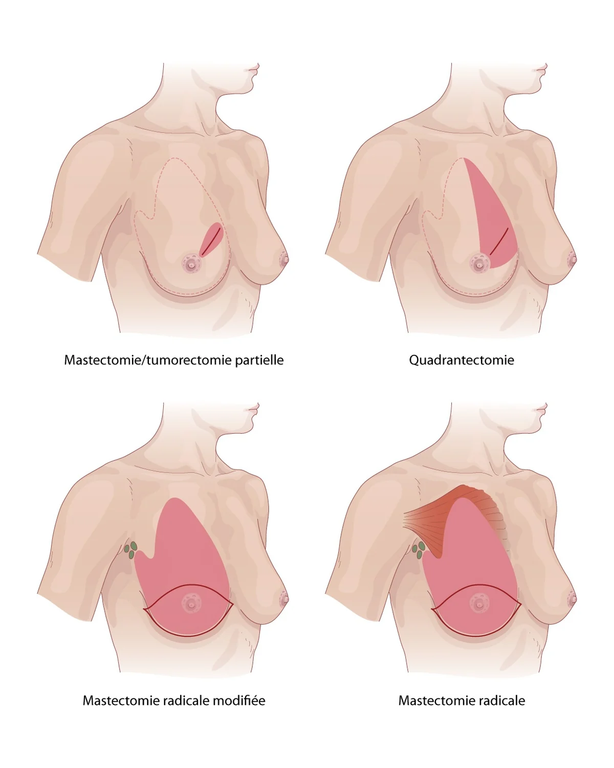
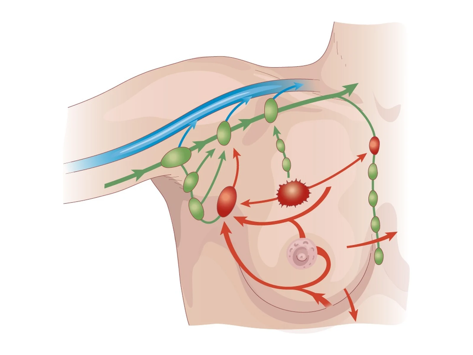
Each case is discussed at a multidisciplinary team meeting, which brings together the surgeon, oncologist, radiation oncologist, radiologist and pathologist. A personalised care plan is then given to you at a dedicated diagnosis consultation.
| Treatment | Aim | For whom? |
|---|---|---|
| Breast-conserving surgery (lumpectomy) | Remove the tumour while keeping the breast | Most small tumours |
| Mastectomy | Remove the whole gland | Large or multiple tumours, patient's choice, contraindication to radiotherapy; reconstruction possible, immediate or delayed |
| Sentinel lymph node biopsy or axillary clearance | Check whether the armpit lymph nodes are involved | Most invasive cancers; sentinel node biopsy limits the risk of lymphoedema (arm swelling) |
| Radiotherapy | Reduce the risk of local recurrence | Almost always after breast-conserving surgery, sometimes after mastectomy |
| Chemotherapy | Treat the disease throughout the body | Tumours at risk of relapse; before surgery (neoadjuvant) or after (adjuvant) |
| Hormone therapy | Block the effect of oestrogen | Hormone receptor-positive tumours, usually for 5 years or more |
| Targeted therapies and immunotherapy | Act on a specific target in the tumour | HER2-positive tumours, some triple-negative tumours or those with a BRCA mutation |
Surgery is performed at the Clinique Ambroise Paré (Neuilly-sur-Seine); the length of hospital stay is explained at the consultation. When treatments may affect fertility, a fertility preservation consultation (freezing eggs or embryos) should be offered to younger women before they start.
What does follow-up after breast cancer treatment involve?
According to the HAS, follow-up combines a clinical examination every 6 months for 2 years, then once a year, and a yearly mammogram, sometimes with an ultrasound, with no time limit. In a patient with no symptoms, repeated CT scans, bone scans and blood markers do not improve survival, so they are not recommended routinely. Follow-up also monitors the effects of hormone therapy (bones, uterus) and supports your return to everyday life. Everything is detailed in our article on follow-up after breast cancer.
When should I see a doctor quickly about a breast change?
Make an appointment within a few days, without waiting for your next screening mammogram, if you notice:
- a new lump in the breast or armpit that persists after your period;
- a recent pulling in of the skin or nipple;
- bloody or clear discharge from one nipple;
- a red, warm, swollen breast when you are not breastfeeding;
- a sore or eczema on the nipple that does not heal.
A red, painful breast with fever during breastfeeding is more likely to be mastitis; it needs a same-day consultation. To book a breast consultation at the practice: appointments can be booked on Doctolib or on 01 43 79 59 79, with an average wait of one week to 10 days, except during holiday periods.
Frequently asked questions
Is breast cancer painful?
Usually not. A cancerous lump is generally painless at first, which is why the absence of pain should not falsely reassure you about a lump that persists.
Should I have a mammogram before 50?
Not routinely. Before 50, a mammogram is prescribed if you have symptoms or an increased risk, such as a family history, after discussion with your doctor.
Can men get breast cancer?
Yes, but it is rare. A lump under the nipple in a man should be examined by a doctor.
Can breast cancer be cured?
Yes, most breast cancers found at an early stage are cured. The outlook depends on the size of the tumour, lymph node involvement and its biological features.
Do I have to pay for breast screening in France?
No. In France, the mammogram and clinical examination done as part of the national screening programme between 50 and 74 are fully covered by the Assurance maladie, with nothing to pay upfront.
References
- Lauby-Secretan B, et al. Breast-cancer screening — viewpoint of the IARC Working Group. N Engl J Med, 2015. doi:10.1056/NEJMsr1504363 — PMID 26039523
- Loibl S, et al. Early breast cancer: ESMO Clinical Practice Guideline for diagnosis, treatment and follow-up. Ann Oncol, 2024. doi:10.1016/j.annonc.2023.11.016 — PMID 38101773
- HAS (French National Authority for Health). Dépistage du cancer du sein en France : identification des femmes à haut risque et modalités de dépistage [Breast cancer screening in France: identifying high-risk women and screening arrangements]. Public health recommendation. 2014. has-sante.fr

Dr Olivier KadochObstetrician-gynaecologist and gynaecological surgeon in Paris (16th and 5th arrondissements). Former chef de clinique-assistant (senior registrar) at the Paris public hospitals (AP-HP); individually accredited by the French National Authority for Health (HAS). RPPS 10003957809. Background and practice locations
A question about your situation?Appointments at the practice or by video consultation, usually within one week to 10 days.
Book an appointmentGeneral information article: it does not replace a medical consultation. In an emergency, call 15 or 112 in France, or your local emergency number.